Confocal Laser Scanning Microscope
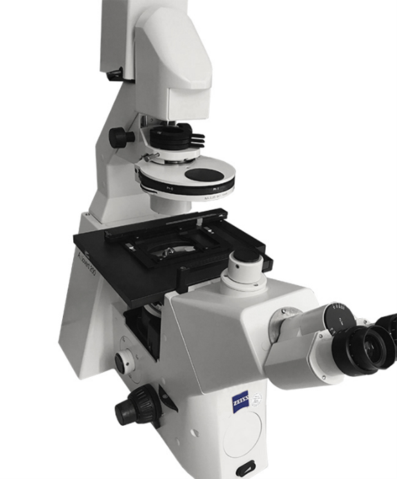
Axiovert 200 M Inverted Research Microscope
Confocal laser scanning microscopy is an advanced optical imaging tool used to obtain high-resolution, three-dimensional images of biological specimens and other materials. The primary feature of the confocal microscope is the ability to eliminate out-of-focus light which results in improved image clarity and contrast that traditional microscopes cannot achieve. Benefits of confocal microscopy include improved resolution, three-dimensional imaging, and reduced photobleaching.
Confocal laser scanning microscopy is an advanced optical imaging tool used to obtain high-resolution, three-dimensional images of biological specimens and other materials. The primary feature of the confocal microscope is the ability to eliminate out-of-focus light which results in improved image clarity and contrast that traditional microscopes cannot achieve. Benefits of confocal microscopy include improved resolution, three-dimensional imaging, and reduced photobleaching.
Resources:
Specifications:
General Methods Description:
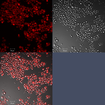
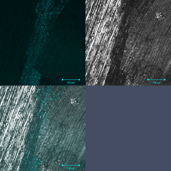
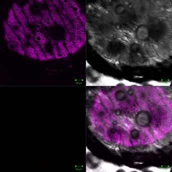
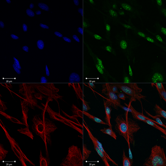
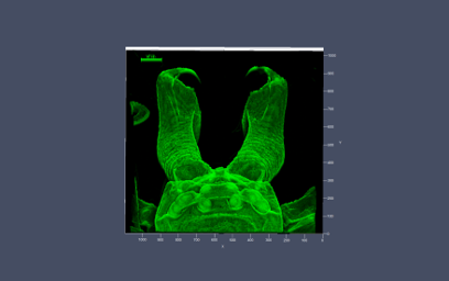
Samples were sectioned, pre-treated with appropriate fluorochromes, and mounted between a glass slide and coverslip using standard antifade media. Fluorescence imaging was performed using a Zeiss LSM 510 confocal laser scanning microscope equipped with excitation laser lines at 405, 458, 477, 488, 514, 543, 594, and 633 nm and matching emission filters for the fluorochromes used. Images were acquired with a high-numerical-aperture objective, and detector settings were optimized to prevent saturation while preserving signal quality.
Specifications:
| Confocal Detection Channels | 3 |
| Laser Lines [nm] | - 405 Diode 30 MW - Argon multiline (458/477/488/514 30 MW) - 543 HeNe 10 MW - 594 HeNe 2 MW - 633 HeNe 5 MW |
| Objectives: "Type" magnification/numerical aperture, Working Distance | - "Plan Apochromat" 5x/0.16, WD=12.1 - EC "Plan Neofluar" 10x/0.30, WD=5.60 - "Plan-Apochromat" 20x/0.80, WD=0.55 - EC Plan "Neofluar" 40x/1.30 Oil DIC, WD=0.20 - "C-Apochromat" 40x/1.20 W corr, WD=0.28 - "Plan-Apochromat" 63x/1.40 Oil DIC, WD=0.18 - "Plan-Apochromat" 100x/1.40 Oil DIC, WD=0.17 |
| Fluorescence Viewing | Mercury light source |
Samples were sectioned, pre-treated with appropriate fluorochromes, and mounted between a glass slide and coverslip using standard antifade media. Fluorescence imaging was performed using a Zeiss LSM 510 confocal laser scanning microscope equipped with excitation laser lines at 405, 458, 477, 488, 514, 543, 594, and 633 nm and matching emission filters for the fluorochromes used. Images were acquired with a high-numerical-aperture objective, and detector settings were optimized to prevent saturation while preserving signal quality.